Dr. Byron Reid, VMD
Whether you’re in the breeding industry, showing at the high-performance level, or riding for pleasure we’ve all experienced the “mare with an attitude” or one with breeding issues.
Today, because of the advancement of medical technology, we are now able to help many owners, riders and breeders ensure their mares have the highest quality of health.
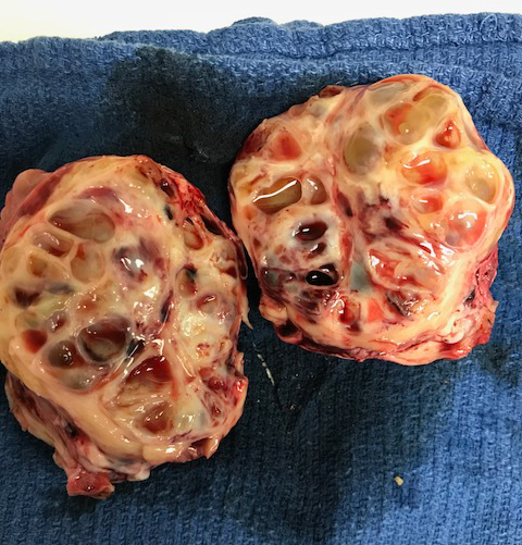
We do some really amazing procedures here at Reid & Associates and one of these involves surgical removal of granulosa-theca cell ovarian tumors. GTC tumors, recognized in mares for centuries, secrete a-number-of hormones which eventually affect a mare’s estrus cycle and often her daily behavior. Some tumors become quite large, and in addition to causing reproductive issues, have been found to cause lameness, recurrent colic episodes, and occasionally stallion-like or bizarre behaviors.
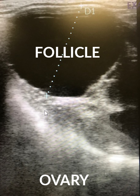
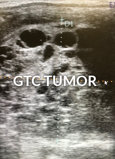
Behavioral or fertility changes have traditionally been the reason for examination and subsequent discovery of these tumors, but recently, with the increasingly common and routine use of reproductive ultrasound examinations, the tumors can be discovered much earlier before behavioral or even fertility issues become present.
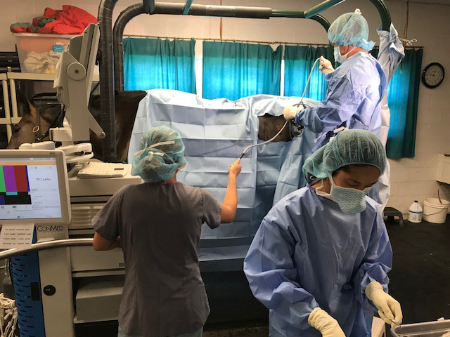
Following discovery of the tumor, surgical removal is indicated. In the past, removal would be done through a large flank incision or through a ventral midline incision under general anesthesia. While these surgical approaches are still occasionally necessary, recently, we are-able-to remove many of these using minimally invasive laparoscopic techniques.
Consequently, the surgery has become much safer and considerably less distressing to the mares. Novel anesthetic techniques and laparoscopic equipment enables us to perform these surgeries through small incisions while the horse is standing—greatly reducing recovery time and improved return to riding and breeding soundness.
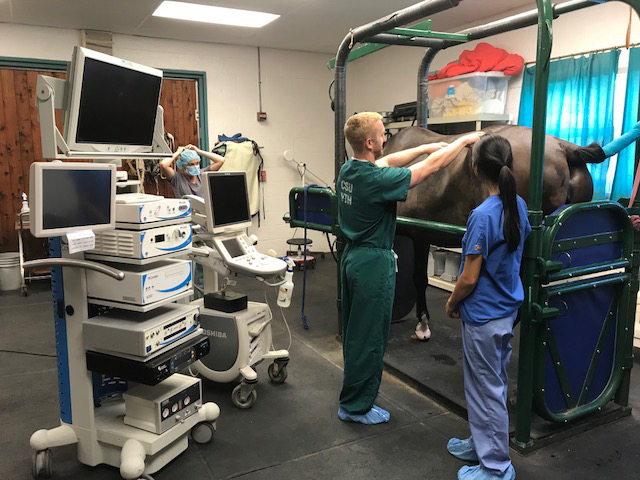
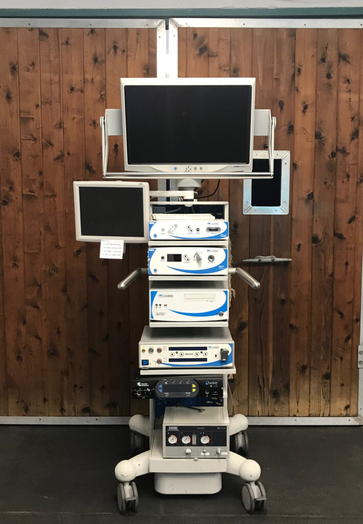
Pictured here is the stack of machines necessary for the surgery. The stack includes a powerful light source attached to a fiberoptic cable that pipes light into the abdomen, a medical grade carbon dioxide tube that pumps carbon dioxide into the abdomen so that we can see between the organs, a high definition camera that projects an image onto a high definition screen, a machine that controls a radio frequency vessel sealing and cutting instrument that ligates and divides the ovarian attachment to the mare’s body, and a morcellator machine that can help us get a large tumor through a small opening by gently breaking it into small pieces inside of a plastic bag.
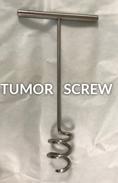
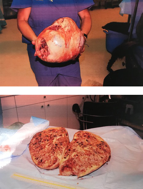
Granulosa Cell Tumors come in many shapes and sizes. Some can be spectacular. In 2008 we had a case that was in Vero Beach and had started to colic. The referring veterinarian had felt a mass and referred the mare. Once the colic had passed we realized that this was a very large ovarian tumor. We planned to remove the tumor through her left flank standing but after laparoscopic severance and ligation of the blood supply, the tumor was so heavy that it could not be lifted through the incision we had made. We therefore closed the incision and took the mare to general anesthesia surgery on her back. We went through a ventral incision identical to a colic exploratory incision and inserted a TUMOR SCREW into the GCT. The tumor screw, (as pictured here) is a very large cork screw looking device that is handy when removing heavy masses. It gets around the problem of getting your two hands AND a tumor through a limited incision. We then closed her up and recovered her. After a very long day for her, she got back to her stall sore and hungry!!!